




















How does a dermatologist examine a mole?
A dermatologist examines a mole by assessing its size, shape, colour, borders, and changes over time, often using dermoscopy to identify features that may require further investigation.
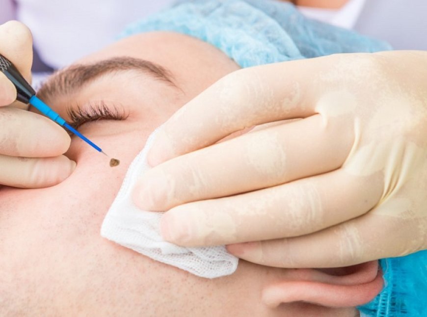
A dermatologist examines a mole by assessing its appearance, history, and changes over time to determine whether it appears harmless or needs further investigation. The examination may involve a close visual inspection, dermoscopy, and, when necessary, a skin biopsy. People considering إزالة الشامات في دبي should have the mole assessed professionally before choosing cosmetic removal. Most moles are harmless, but a new, changing, itching, or bleeding mole may require closer evaluation.
What Happens During a Mole Examination?
The examination usually begins with a discussion about the mole and the patient's medical history. The dermatologist may ask when the mole first appeared, whether it has changed, and whether it has caused symptoms such as itching, bleeding, tenderness, or irritation.
The dermatologist may also ask about previous skin problems, family history of melanoma or other skin cancers, previous mole removals, and the patient's general skin health. This information helps provide context for what the dermatologist sees during the physical examination.
What Does a Dermatologist Look For?
During the examination, the dermatologist carefully evaluates the mole and the surrounding skin. A typical harmless mole is often one colour and round or oval, although normal moles can vary considerably in appearance. The dermatologist looks for features that make a mole different from the patient's other moles or suggest that it may require further investigation.
Important features include:
- Size and diameter
- Shape and symmetry
- Border definition
- Colour and colour variation
- Surface and texture
- Whether the mole is flat or raised
- Changes in appearance
- Bleeding, itching, or other symptoms
A mole that looks different from the surrounding moles or has changed over time deserves particular attention.
What Is the ABCDE Method?
Dermatologists commonly use the ABCDE approach when assessing pigmented lesions for possible melanoma warning signs.
A – Asymmetry: One half of the mole looks different from the other.
B – Border: The border is irregular, scalloped, uneven, or poorly defined.
C – Colour: The mole contains different colours or areas of changing pigmentation.
D – Diameter: The lesion is larger than approximately 6 millimetres, although melanoma can be smaller.
E – Evolving: The mole changes in size, shape, colour, or overall appearance.
These signs do not automatically mean that a mole is cancerous. However, they can indicate that professional assessment is needed.
What Is Dermoscopy?
Dermoscopy is a close examination of the skin using a specialised handheld device called a dermatoscope. It provides magnification and helps the dermatologist see structures and pigmentation patterns that may not be visible during an ordinary visual examination.
The dermatologist may use dermoscopy to assess whether a mole has characteristics associated with a benign lesion or whether additional investigation is appropriate.
Dermoscopy does not replace a biopsy when cancer is suspected. Instead, it can help the dermatologist decide which lesions require further examination.
Does the Dermatologist Examine the Rest of the Skin?
It depends on the reason for the appointment. If a patient is concerned about one particular mole, the dermatologist may focus on that lesion and the surrounding skin. If there are multiple concerning spots or the patient has risk factors for skin cancer, a broader skin examination may be recommended.
A full-body skin examination can include the scalp, face, neck, trunk, arms, legs, hands, feet, nails, and other areas where skin cancer can develop. The dermatologist may compare different moles to identify a lesion that looks noticeably different from the patient's other spots.
When Is a Mole Biopsy Necessary?
Not every mole needs a biopsy. If a dermatologist sees warning signs that could indicate skin cancer, they may recommend removing part or all of the mole for laboratory examination.
During a skin biopsy, the area is usually numbed with local anaesthetic. The dermatologist removes a sample of the suspicious lesion and sends it to a laboratory, where it is examined under a microscope.
Microscopic examination is the only way to confirm whether a lesion contains skin cancer. The biopsy results can also identify the specific type of skin condition present.
Can a Mole Be Removed During the Same Visit?
In many cases, a dermatologist can remove a mole during an office visit if it is considered appropriate. The American Academy of Dermatology explains that most mole removals require only one office visit, although some patients may need a second appointment.
Common removal techniques include surgical shave removal and surgical excision. The selected method depends on the mole's characteristics and whether the tissue needs to be examined.
If the mole is suspicious, the priority is appropriate diagnosis rather than simply removing it for cosmetic reasons.
What If the Mole Looks Completely Normal?
A stable mole that has no concerning features may not need to be removed. Dermatologists generally do not recommend removing every mole because many are harmless and can simply be monitored.
Removal may still be considered if a benign mole repeatedly catches on clothing, becomes irritated during shaving, or is cosmetically unwanted.
The dermatologist can discuss whether removal is appropriate and explain the possible benefits and risks.
What Should You Tell the Dermatologist?
Before the examination, it is useful to explain any changes you have noticed. Tell the dermatologist if the mole has:
- Increased in size
- Changed shape
- Developed new colours
- Become itchy
- Started bleeding
- Become painful
- Developed a crust or sore
- Changed differently from your other moles
You should also mention any previous skin cancer diagnosis, family history of melanoma, previous mole removal, and relevant medications or medical conditions.
Providing an accurate history can help the dermatologist decide whether monitoring, removal, or biopsy is appropriate.
Why Should You Avoid Removing a Mole Before Examination?
Moles should not be cut, burned, shaved, or treated with unapproved home-removal products. Attempting removal at home can cause infection and scarring, and it may also delay the diagnosis of skin cancer if the lesion is suspicious.
Professional assessment is particularly important for a mole that is changing, itching, bleeding, or different from the surrounding moles. Early evaluation can help identify concerning lesions before they progress.
Frequently Asked Questions:
Does examining a mole hurt?
A routine visual examination and dermoscopy are generally non-invasive. If a biopsy is required, the area is usually numbed with local anaesthetic before tissue is removed.
Can a dermatologist tell if a mole is cancerous just by looking at it?
A dermatologist can identify features that are concerning, but visual examination alone cannot definitively diagnose skin cancer. A biopsy and microscopic examination are required when cancer is suspected.
Do all suspicious moles need to be removed?
A suspicious mole may need a biopsy or complete removal depending on the clinical assessment. The dermatologist determines the appropriate approach.
Should a new mole be examined?
A new mole, particularly one that appears during adulthood, should be assessed if it is changing or has unusual features. The AAD recommends professional evaluation for new or changing moles and spots that itch or bleed.
Can a dermatologist examine several moles at once?
Yes. During a broader skin examination, a dermatologist can assess multiple moles and other skin spots and identify those that may require closer monitoring or investigation.
Conclusion:
A dermatologist examines a mole by reviewing its history, carefully inspecting its size, shape, border, colour, and evolution, and using dermoscopy when appropriate. If the mole has suspicious characteristics, a biopsy may be recommended so the tissue can be examined under a microscope. Stable and harmless moles may simply be monitored, while bothersome or medically concerning moles can be considered for professional removal. Any mole that changes, itches, bleeds, or looks different from other moles should receive timely dermatological assessment.
What's Your Reaction?
 Like
0
 Dislike
0
 Love
0
 Funny
0
 Angry
0
 Sad
0
 Wow
0