


Post-Procedural Complications: Managing Infections and Keloids in Riyadh Patients.
Post-Procedural Complications: Managing Infections and Keloids in Riyadh Patients
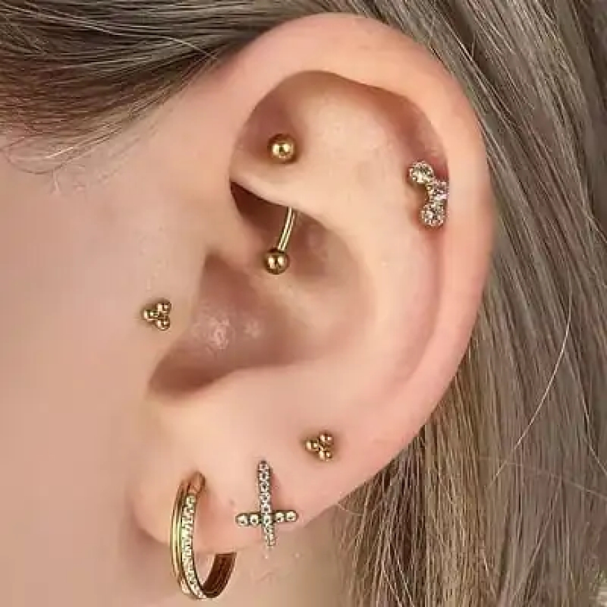
Ear Piercing in Riyadh has become a significant aesthetic interest, yet the popularity of the procedure is often accompanied by an increase in post-procedural complications. Managing these issues—specifically bacterial infections and abnormal scar formation—requires a structured, medically-sound approach. When a piercing site fails to heal correctly, early intervention by qualified healthcare professionals is critical to prevent the progression of localized tissue issues into systemic complications or permanent disfigurement.
Clinical Identification of Infectious Complications
The clinical presentation of an infected piercing often begins with localized erythema (redness), edema (swelling), and tenderness. However, differentiating between a standard healing response and an active infection is essential. In a medical context, signs such as persistent or worsening pain, an increase in local temperature, and the presence of purulent discharge (pus) are indicators that the body’s natural immune response has been overwhelmed by bacterial colonization.
In the context of the ear, cartilage piercings present a particularly high risk due to the tissue's limited blood supply. Infections here, such as auricular chondritis, are frequently linked to Pseudomonas aeruginosa, a pathogen that can lead to rapid cartilage necrosis. In Riyadh’s clinical practice, managing these cases often requires empirical antibiotic therapy that provides broad-spectrum coverage, specifically targeting Pseudomonas and common staphylococcal strains. It is generally advised not to remove the jewelry during the initial management phase, as the closure of the wound may trap the infection and lead to the formation of an abscess.
Differentiating Hypertrophic Scars from Keloids
One of the most frequent non-infectious complications encountered by patients is the development of raised tissue at the site of the piercing. It is vital to distinguish between hypertrophic scars and true keloids:
-
Hypertrophic Scars: These are localized, raised bumps that remain confined to the original injury site. They often occur during the first few weeks or months of the healing process and may regress over time with proper maintenance, such as minimizing mechanical irritation and using silicone-based topical treatments.
-
Keloids: These represent an abnormal, excessive production of collagen. Unlike hypertrophic scars, keloids extend beyond the borders of the initial piercing site and can continue to proliferate. They are characterized by their firm, rubbery texture and may cause pruritus (itching) or tenderness.
Genetic predisposition and skin type are significant factors in keloid formation. For patients with a history of abnormal scarring, elective piercings in high-risk zones—such as the pinna or the cartilaginous regions of the ear—should be carefully evaluated, as the likelihood of recurrence is high.
Multi-Modal Management Strategies
In clinical settings, the management of these complications follows a hierarchical strategy based on the severity and nature of the lesion. For early-stage scar management, non-invasive methods are prioritized. This includes:
-
Pressure Therapy: The use of customized, low-level compression devices or clips to influence collagen remodeling through localized pressure.
-
Topical Interventions: The application of medical-grade silicone gel sheets or creams, which have been shown to flatten scars by hydrating the tissue and modulating the inflammatory response.
When conservative measures are insufficient, especially in the case of mature keloids, medical intervention is required. Treatments often include:
-
Intralesional Corticosteroid Injections: These are utilized to break down collagen bundles and reduce localized inflammation.
-
Cryotherapy and Laser Therapy: These methods are employed to reduce the volume and vascularity of the scar tissue.
-
Surgical Excision: Often reserved for large, resistant keloids, surgical removal is typically combined with adjunct therapies (such as pressure therapy or post-operative monitoring) to minimize the high risk of recurrence.
The Importance of Proactive Aftercare
Prevention remains the most effective management strategy. Patients are encouraged to adhere to a strict hygiene regimen, which involves cleaning the area twice daily with a sterile saline solution while avoiding harsh agents like alcohol or hydrogen peroxide, which can desiccate the tissue and impede the healing process. Furthermore, minimizing physical trauma—such as twisting the jewelry or allowing the piercing to be exposed to friction from hair or clothing—is fundamental to reducing the inflammatory triggers that lead to both infection and abnormal scar proliferation. By fostering a clinical partnership that prioritizes long-term anatomical integrity, patients can successfully navigate the healing period and minimize the impact of post-procedural complications.
What's Your Reaction?
 Like
0
Like
0
 Dislike
0
Dislike
0
 Love
0
Love
0
 Funny
0
Funny
0
 Angry
0
Angry
0
 Sad
0
Sad
0
 Wow
0
Wow
0