


ELISA Assay Method in COVID-19 Antibody Detection: What You Should Know
Learn how the ELISA assay method detects COVID-19 antibodies. Understand its laboratory workflow, diagnostic value, and role in tracking immunity.
The COVID-19 pandemic highlighted the need for robust diagnostic tools to detect infections and understand immune responses. While PCR tests identified active infections, ELISA became essential for serological testing, detecting antibodies against SARS-CoV-2. This article explores how ELISA works, its role in antibody detection, and its comparison with advanced techniques such as LC-MS.
The Science Behind ELISA
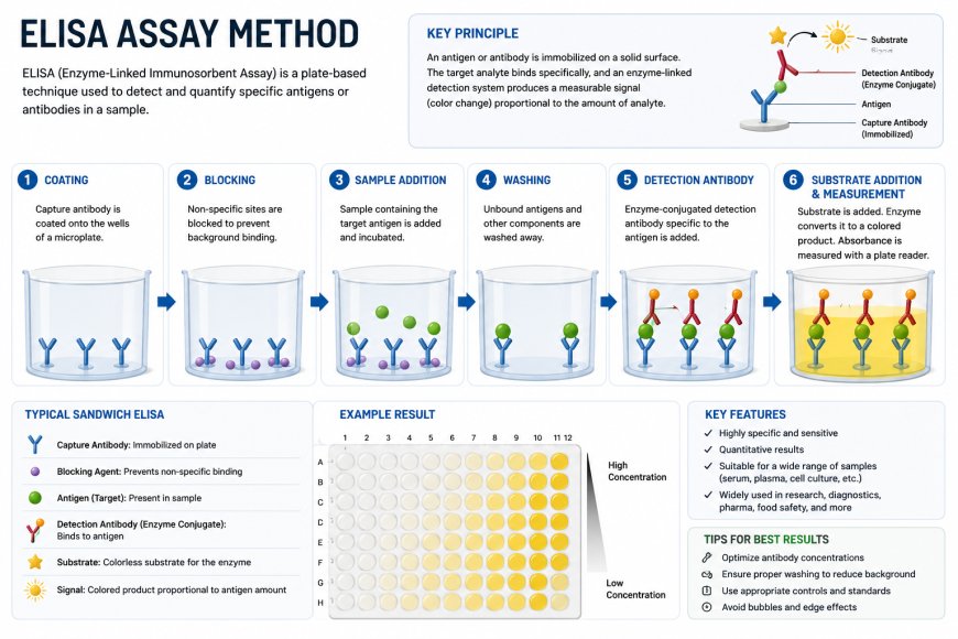
The ELISA immunoassay relies on the specific binding between an antigen and an antibody. It uses an enzyme-linked conjugate and a substrate to generate a measurable signal, such as a color change, that is proportional to the amount of analyte present.
For COVID-19 testing, ELISA detects host antibodies (IgG, IgM, IgA) against viral antigens such as the Spike (S) or Nucleocapsid (N) protein. The main components are:
- Solid Phase: A microplate coated with a SARS-CoV-2 antigen.
- Sample: Patient serum or plasma.
- Conjugate: An enzyme-labeled secondary antibody.
- Substrate: A chemical that reacts with the enzyme to create a signal.
When a patient's sample is added, specific antibodies bind to the antigen. Washing steps remove unbound materials, ensuring the resulting signal is specific to the target antibody.
ELISA Assay Procedure: A Step-by-Step Guide
Executing an ELISA analysis requires precision and adherence to a strict protocol to ensure reproducibility. While variations exist (direct, indirect, sandwich, competitive), the indirect ELISA is commonly used for antibody detection.
- Coating: The microplate wells are coated with the viral antigen (e.g., recombinant SARS-CoV-2 protein) and incubated overnight.
- Blocking: A blocking buffer containing non-reactive proteins (like BSA) is added to cover any remaining binding sites on the plate surface. This prevents non-specific binding of the patient antibodies.
- Sample Incubation: Patient serum is diluted and added to the wells. If SARS-CoV-2 antibodies are present, they bind to the coated antigen.
- Washing: The plate is washed with a specific buffer to remove unbound antibodies and other serum components.
- Detection: An enzyme-conjugated secondary antibody (e.g., anti-human IgG linked to Horseradish Peroxidase) is added. This antibody binds to the patient's antibodies that are captured on the plate.
- Substrate Addition: A substrate solution (such as TMB) is added. The enzyme on the secondary antibody converts the substrate into a colored product.
- Stop Solution: An acidic solution is added to stop the enzymatic reaction.
- Measurement: The optical density (OD) is measured using a spectrophotometer. The color intensity is directly proportional to the concentration of antibodies in the sample.
Proper ELISA validation is required to establish the assay's sensitivity, specificity, and range, ensuring reliable results for clinical or research use.
Must Read: Immunogenicity Assay Role in Pharmacokinetics Assay Studies
Advantages and Limitations of ELISA in Antibody Detection
ELISA remains a staple in immunology due to its established track record and accessibility. However, like any analytical method, it has distinct pros and cons.
Advantages
- High Sensitivity and Specificity: When properly optimized, ELISA can detect low levels of antibodies with high accuracy.
- High Throughput: The 96-well plate format allows for the simultaneous ELISA analysis of many samples, making it efficient for population screening.
- Quantitative Capabilities: It provides quantitative data regarding antibody titers, rather than just a binary positive/negative result.
- Simplicity: The equipment required is standard in most ELISA labs, making it accessible for widespread use.
Limitations
- Cross-Reactivity: Antibodies against other coronaviruses may bind to the antigen, leading to false positives.
- Labor Intensive: Traditional methods require multiple manual steps, increasing the risk of human error.
- Limited Multiplexing: Standard ELISA typically detects one analyte per well. While multiplexed ELISA formats exist, they are often more complex to optimize than single-plex assays.
LC MS MS Analysis: A Comparison with ELISA
Liquid Chromatography with Tandem Mass Spectrometry (LC-MS/MS) provides an alternative to ELISA, relying on mass analysis rather than antibody-antigen interactions. It separates molecules based on their physicochemical properties and identifies them by their mass-to-charge ratio.
Key Differences from ELISA
- Specificity: LC-MS/MS offers superior specificity by directly detecting specific peptides from the target protein, minimizing the risk of interference from non-specific binding common in ELISA.
- Development Time: LC-MS methods can be developed more rapidly than ELISA immunoassays, as synthetic peptides are easier to generate than the high-quality antibodies required for ELISA.
- Cost and Complexity: ELISA is more cost-effective and easier to use for routine applications. In contrast, LC-MS/MS requires specialized equipment and personnel, making it better suited for validation and reference testing rather than high-throughput screening.
The Future of Antibody Testing
The COVID-19 pandemic accelerated advancements in diagnostic technologies. While ELISA is a primary tool for serological testing due to its high throughput and cost-effectiveness in Assay Testing, mass spectrometry provides superior specificity, which is vital for advanced diagnostics.
The future of antibody testing likely involves a hybrid approach. Multiplexed ELISA platforms could enable simultaneous screening for multiple variants, while LC-MS could be utilized for biomarker discovery and validating immunoassays. This combination of methodologies will enhance surveillance and deepen our understanding of infectious diseases.
What's Your Reaction?
 Like
0
Like
0
 Dislike
0
Dislike
0
 Love
0
Love
0
 Funny
0
Funny
0
 Angry
0
Angry
0
 Sad
0
Sad
0
 Wow
0
Wow
0